TERRITORIAL FIGHTING WITH FATAL CONSEQUENCES: ROE DEER WITH SKULL AND BRAIN DAMAGE
- Wildlife Health Ghent

- May 23, 2025
- 2 min read
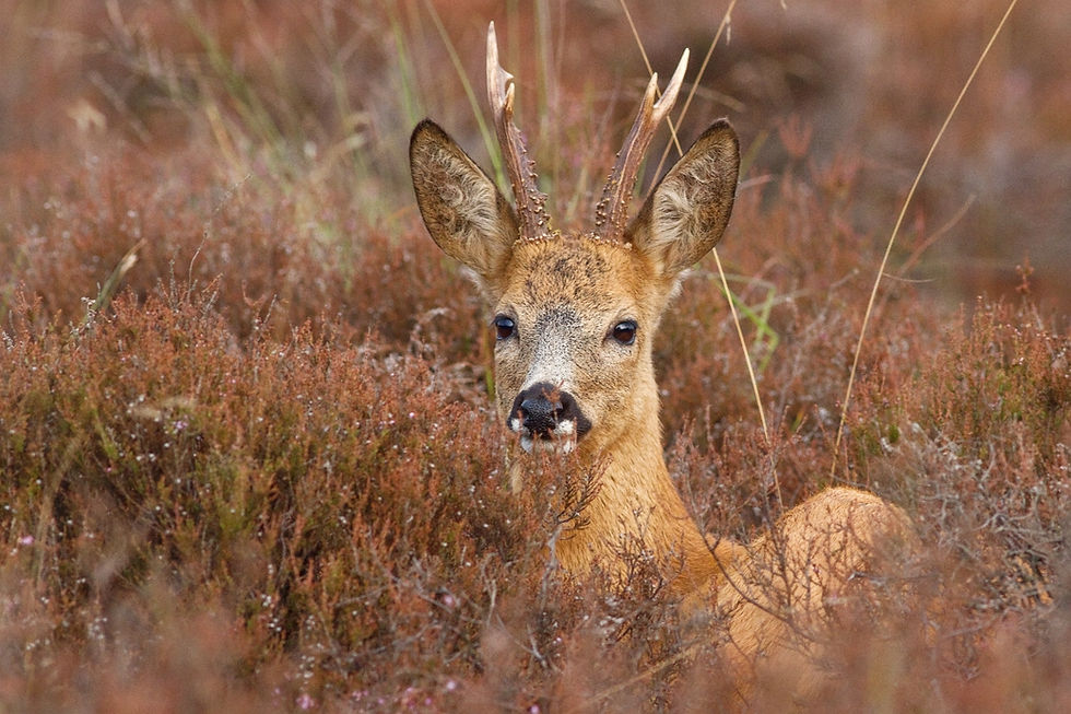
During spring and summer, roe deer ( Capreolus capreolus ) display pronounced territorial behavior. Fights between males are frequent and can lead to injuries. In some cases, these injuries cause serious complications, as shown by two cases reported via the wildlife disease surveillance program of the Agency for Nature and Forests (ANB).
CASE 1
In June 2024, a roe deer was brought in from the Geraardsbergen region. The animal showed abnormal behavior and could easily be approached. It walked in circles - a neurological alarm signal. During the capture, the animal still reacted alertly, but due to the poor prognosis, it was decided to euthanize him.
At necropsy, a small, foul-smelling wound was noted at the base of the antlers, with the presence of maggots. Although the wound appeared superficial at first glance, further inspection revealed that the injury extended deeper. A cross-section of the skull revealed necrotic bone tissue due to osteomyelitis . The initial injury was likely caused by an antler impact from another buck. Such deep puncture wounds provide a portal of entry for bacteria to travel along bone sutures and into the brain.


The animal developed suppurative meningoencephalitis, a purulent inflammation of both the meninges and brain tissue. Histological examination showed perivascular inflammatory infiltrates with neutrophils, lymphocytes, plasma cells and macrophages, as well as small areas of gliosis – a sign of damage to the nervous tissue.

CASE 2
A more recent case occurred last week, when a debilitated buck was euthanized after persistent symptoms of lethargy and excessive salivation, despite administration of supportive medication. Upon necropsy, loose antlers were noted. After sawing off the base of the antlers and sawing through the skull, pus accumulation was found, both around the antlers and at the base of the antlers. Purulent substances were also found on the surface of the cerebrum, indicating that the bacterial infection had already broken through to the central nervous system. Bacteriological examination of this identified the bacterium Trueperella pyogenes as the culprit.


These cases are not unique to Flanders. Similar injuries have been described in several European countries, mainly in regions with a high buck-doe ratio (Pewsner et al., 2017; Cohen et al., 2015). In such areas, the frequency and intensity of fights increases, which increases the risk of deep wounds and secondary infections. These cases illustrate how intraspecific behaviour in roe deer can, under certain circumstances, lead to serious neurological disorders with fatal outcome.


Comments