UNCOMMON JAW TUMOR WITH BONE INFECTION IN ROE DEER
- Wildlife Health Ghent

- Jun 30, 2025
- 3 min read
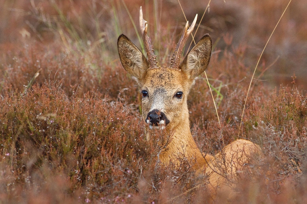
In the spring of 2025, an adult roe deer (Capreolus capreolus) was found dead and submitted to Wildlife Health Ghent for examination through the disease surveillance program of the Agency for Nature and Forests (ANB).
The animal was severely emaciated and dehydrated, with a prominent growth (5 x 3 cm) on the right lower jaw. This mass had displaced several teeth and had caused visible bone damage (osteolysis), along with open ulcers and inflammation. No signs of cancer spread (metastasis) were found in other organs. However, the lesion caused serious disfigurement and a chronic bone infection (osteomyelitis), which likely contributed to the animal’s death.


How Common Are Jaw Tumors in Deer?
Jaw masses caused by infection (such as those from Trueperella pyogenes or Actinomyces bovis) are relatively common in herbivores, but true tumors in wild ruminants' mouths are exceptionally rare. Some unusual examples exist:
A cementoblastoma found in a red deer from the Late Pleistocene in France (Kierdorf et al., 2015)
A composite odontoma (a rare benign growth) in a malformed incisor of a red deer from the French Alps (Binois et al., 2014)
An odontoameloblastoma in a free-ranging red deer that had to be euthanized due to poor condition and a large jaw mass (Hartung et al., 2021)
The roe deer is the most widespread deer species in Europe, but detailed reports on its diseases are still limited. For example, a large study in Switzerland covering 1571 roe deer necropsies between 1958 and 2014 reported only ten cases involving tumours in the head, including a fibrosarcoma—but without clear detail on its location (Pewsner et al., 2017).
What Did We See in This Case?
Histological (microscopic) analysis revealed a highly cellular, infiltrative mass made of spindle-shaped cells arranged in bundles, with mild variation in cell size and rare cell division (mitoses). Several necrotic (dead) zones were present, and the tumour surrounded fragments of bone. It extended to the overlying gum tissue, which was ulcerated. The surrounding bone had signs of chronic inflammation, with fibrous tissue, activated bone cells (osteoblasts and osteoclasts), and infection. Based on this, the lesion was most compatible with a mandibular fibrosarcoma with secondary osteomyelitis. A spindle-cell type osteosarcoma was considered less likely but could not be fully ruled out.

How Rare Is This?
Oral fibrosarcoma is a malignant connective tissue tumor seen relatively often in veterinary practice, especially in middle-aged, medium to large dogs. But in deer, it's an extremely rare finding. Only one prior case was found in the literature: a fibrosarcoma in a male Père David’s Deer that developed a similar jaw mass and had to be euthanized (Hubbard et al., 1983).
Take-Home Message
This case illustrates a rare occurrence of a jaw tumor in a wild roe deer, complicated by a secondary infection. It highlights the importance of wildlife disease surveillance in detecting unusual or emerging health issues in native species.


Comments